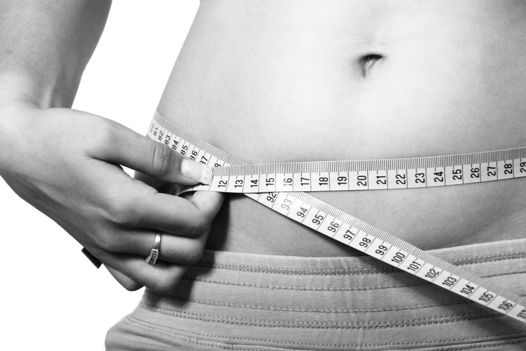
From Butts, Guts, and Other Problem Areas:
Grasso gordo gruby gras
Have you ever wondered what causes stubborn fat? How exactly does that fast food hamburger, side order of French fries, Strawberry milkshake, and apple pie served in cardboard cutout make us fat?
Physiologically speaking, what actions go on inside our body after we have eaten the last spoonful of Frusen Gladja that so readily plasters deposits of unsightly fat around our midsection or on our thighs. In other words, how does what we put in our mouth turn to the fat on our buns, above our hips, below our chins, and around our bellies? And…which part of the body is the hardest to lose fat and can you lose stubborn fat?

The term fat, derived from the Latin word obesus, was used to describe individuals whose health was suffering due to overindulgence. Fat refers to fat cells known as adipocytes, that are stored in different areas throughout our body. There are three main categories of fat found in the human body: visceral fat, subcutaneous fat, and brown adipose tissue (BAT). Each of these serves a different function in the body.
Fat is primarily stored in designated fat storage cells called adipocytes. Adipocytes are stored in different areas throughout the body, primarily under the skin (subcutaneous) or surrounding vital organs (visceral fat) for protection.
In several research studies, fat tissue has been found to be an endocrine tissue. Researchers have identified nearly eighty different proteins that are secreted by fat cells (Rosenow). Some of the major hormones secreted by fat tissue include leptin, resistin, and estrogen, all of which can play an active role in the development of obesity.
Leptin is a satiety hormone that signals the brain to let us know when we are full. Resistin is a pro-inflammatory hormone that may play a role in insulin resistance, and estrogen has been associated with various diseases including cancers, Parkinson’s Disease, and obesity.
TYPES OF fAT
Brown Adipose Tissue
Metabolically active type of fat that is responsible for thermoregulation as it turns food into heat. Has more capillaries than white fat. High mitochondria content gives BAT it’s brown coloration.
Neutral Colored Adipose Tissue (brite)
White fat tissue that expresses thermogenic capacity.
White Subcutaneous Fat
Most common type of fat in the body, this metabolically inactive fat directly under the skin, found about the hips, thighs, belly, and butt (think cellulite). Subcutaneous fat produces the hormone adiponectin, a hormone that plays an important role in glucose regulation and lipid metabolism in insulin sensitive tissues. The larger the fat cells, the less adiponectin you produce, potentially leading to metabolic disorders. The smaller the fat cells, the more adiponectin produced.
White Visceral Fat
The deep fat that surrounds our internal organs, visceral fat releases immune system substances called cytokines that can affect insulin production and promote chronic inflammation. Visceral fat also plays a role in the development of cholesterol as it empties toxins, metabolites, and fatty acids into the bloodstream and liver (8).
Cellulite
Cellulite is the uneven, dimpled/marbled looking fat that often rears its head about the butt, thighs, and midsection. It has been referred to as appearing like cottage cheese or an orange peel. Our fat cells are stored in compartments just below the dermis. This layer of subcutaneous fat is just above a layer of connective tissue that is housed between the fat and muscle tissue.
When the stability of the connective collagen tissue layer is weakened due to reasons including hormonal changes, poor circulation, lack of exercise, hydration issues, smoking, poor nutrition, age, gender, estrogenic toxins, and metabolic changes, hard fibrous bands may form in the connective tissue. The subcutaneous fat is then compressed, leading to herniation. This is when the “cottage cheese/orange peel” appearance presents (19).
There are gender differences in this collagen tissue that increase the risk of cellulite appearance. Men may have a thicker dermis layer that increases the risk of internal expansion, while women run the risk of external expansion leading to an increased risk of cellulite appearance (Rosenbaum 1998). The good news is that females who lose weight seem to make the appearance of cellulite disappear.
Hormones and Fat Storage Areas in the Body
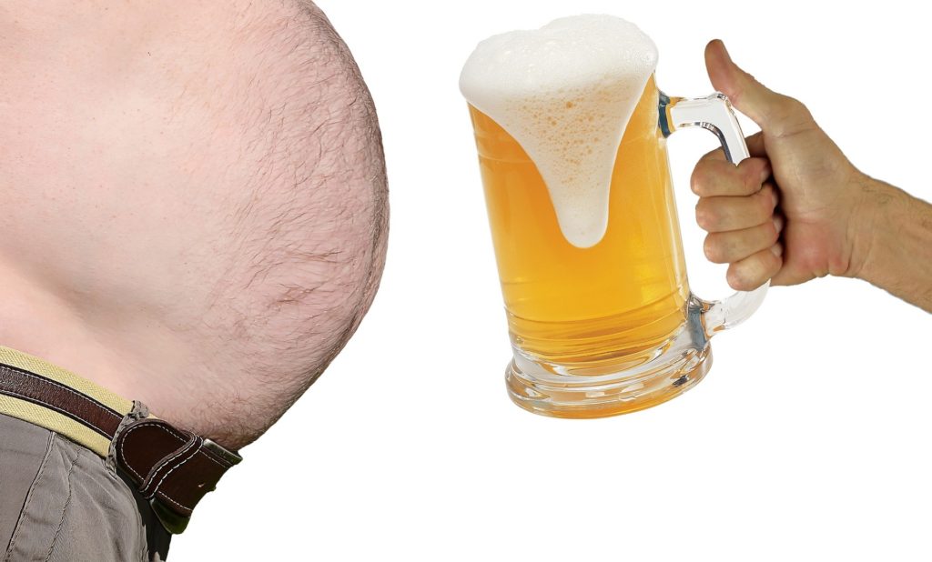
Did you know that your hormones play a critical role in where you are storing your body fat? The late research scientist Per Bjorntorp is quite possibly the most significant contributor to the science of hormonal control and regulation of body fat storage.
In some of his earlier works, Bjorntorp was already beginning to correlate fat distribution patterns and their hormonal regulation. Noticing deficiencies in certain anabolic hormones or excess secretion of storage and/or catabolic hormones led to fat distribution patterns in men and women. From his early research on serum androgen levels and fat distribution in women to his studies on testosterone replacement in men with excess belly fat, Bjorntorp’s work has played a critical role in the science of hormones and fat distribution.
Truly a pioneer in this field, Bjorntorp began seeing similarities in fat distribution patterning and hormonal profiles of his subjects as early as the late 80’s early 90’s. In 1991, the International Journal of Obesity printed his research paper “Adipose tissue distribution and function”, in which Bjorntorp provided some magnificent insight into the hormonal regulation of fat storage. Excesses or deficiencies in certain hormones could lead to fat accumulation in different areas of the human body.
Many of Bjorntorp’s other works were published in the pages of other highly respected journals, from the aptly named “Hormonal control of regional fat destruction (1997)” found in the pages of the Human Reproduction publication, to his works in Hormonal Research and the International Journal of Obesity including “Endocrine-metabolic pattern and adipose tissue distribution (1993)”, “Adipose tissue distribution and function (1991)”, and “The regulation of adipose tissue distribution in humans (1996)”, Bjorntorp’s work was truly cutting edge.
The other trailblazer in the field of hormones and fat distribution patterning was the late, great, mentor to thousands, Charles Poliquin. HIs biosignature modulation was and is still to this day a revolutionary approach to regional fat patterning and how to modulate it. .
How do some of your major hormones affect weight gain? If highly insulinogenic chemical laden processed foods make up the majority of your diet, the hormone insulin may be compromised. More may be required, leading to a decrease in its action, efficiency, and sensitivity. If you are stressed, whether physically (poor nutrition can be stressful to the body) or emotionally, your body may increase the production of the stress hormone cortisol, which signals the body to go into storage mode. Storage of calories and eventually fat accumulation can result.
If your thyroid is not functioning optimally, your metabolism may slow down, which in turn may lead to weight gain. If your testosterone levels are similar to those seen in pygmy goats, you may see a dramatic in increase in body fat accumulation due to the decreases in lean muscle tissue. With a decrease in testosterone, you may see an increase in the “female” hormone estrogen, which is conveniently stored in fat cells. With more estrogen, you will need more and larger fat cells to accommodate.
Other hormones such as insulin, GH, the catecholamines, glucagon, serotonin, and neuropeptide Y are under the regulation of other glands such as the adrenals and brain. Fat cells have even been shown to be endocrine tissue themselves, with the production of hormones such as leptin and ghrelin.
Apples and Pears

A study published in The Journal of American Medical Association looked at how people of different body fat distribution patterns, apple or pear, respond to different types of diets. The goal was to use two different diets that affected insulin sensitivity and see how these diets performed in subjects with different fat distribution patterns. The diets were low carb and low fat. Understanding that the apple fat distribution pattern subjects secrete more insulin post meal and that the pears secreted less. The apples lost more weight on the low carb diet and were able to maintain for 18 months, while the pears lost weight on both diets as long as they had a reduced caloric intake. The downside of being a pear was that the weight came off more slowly and was also gained more quickly (4).
Did you know that where you store your fat has been linked to cardiovascular disease risk factors?
A 1989 study from The American Journal of Clinical Nutrition found both the ratio of triceps and subscapular skinfold measurements and waist to hip girths were associated with higher concentrations of serum triglycerides and lower levels of good HDL cholesterol (9).
In keeping with the CVD risk factors and body fat storage patterns a separate, much earlier study from The American Journal of Clinical Nutrition, looked at the reliability of the triceps skinfold thickness versus the subscapular skinfold thickness as a measure of overall obesity.
The researchers found that in obese women, the triceps skinfold measurement was greater than the subscapular measurement in 83% of those women tested. The subscapular measurement was greater in only 12% of the subjects. The researchers stated “We were struck by the fact that in a number of particularly obese women there was a notable absence of a marked excess of fat in the region of the back (subscapular) while at the same time in all instances the thickness of the triceps skin fold indicated marked excessive adipose tissue. Thus, there appear to be obese women who have excessive fat deposits on the upper arm but not on the back (22).”
Another earlier study from The American Journal of Clinical Nutrition looked at the relationships between skinfold thickness and blood sugar/blood fats in normal men. The researchers found the “highest triglyceride levels were found in men who were moderately obese and had acquired their excess adiposity during adult life. The thickness of the scapular and costal skinfolds was strongly correlated with the other measures of fatness (1).”
An interesting finding of the study was the correlation between forearm thickness and metabolic disorders, in particular high serum triglycerides. The researchers expressed “It is apparent that the thinner the forearm, the more strongly is central adiposity correlated with serum triglyceride concentration. The man whose innate state of thinness is indicated by a thin forearm could thus become obese only at the cost of overloading his existing centrally located adipose cells, while the naturally obese man, generously endowed with adipose cells, could easily expand his adipose tissue without overloading any one cell. The fact that the mean triglyceride levels in the innately thin man who becomes obese are higher than in the innately obese man suggests that hyperglyceridemia may result from overloading of existing adipose cells. This hypothesis would account for the frequently normal triglyceride concentrations in very obese men and the high triglyceride concentrations in thin men who have gained even a modest amount of weight (1).”
“The correlation between weight gain during adult life and fat thickness of the trunk but not of the extremities lends some support to the hypothesis that there are two types of obesity: 1) hereditary obesity, or obesity acquired very early in life, in which adiposity is generalized and involves the extremities as well as the trunk; and 2) acquired, or perhaps cultural obesity resulting from overeating and underactivity after maturity in which obesity is largely of the trunk and not extremities (1).”
The researchers concluded “that there are two kinds of fat men: those with thin forearms who are by nature thin but who have gained weight during adult life, and those with fat forearms who have been fat all their lives. Abdominal skinfold thickness greater than 20mm is associated with serum triglyceride levels that are markedly higher in naturally thin men than naturally fat men (1).”
Impact of Training on Fat Patterning

Can the type of training you do affect where you lose fat from? In a 2005 study from The British Journal of Sports Medicine, researchers followed 24 male and 13 female runners who engaged in intense training over a three-year period. The runners were classified into groups according to their best performance capabilities according to event; sprint trained consisted of 100m and 400m; endurance trained consisted of 800m, 1500m, 3000, 5000m, 10,000m, and marathon.
To be included in this study, athletes were required to compete at a national or international level for at least two years. 12 of the males and 8 of the females were Olympic athletes. All athletes trained six or seven days a week (20-25hrs) during the season.
Skinfold thicknesses were taken at the biceps, triceps, subscapular, pectoral, iliac crest, abdominal, front thigh, and medial calf. After one, two, and three years of training, there was a significant increase in performance and decreases in the sum of six skinfolds. There were no significant differences in body weight, triceps, subscapular, pectoral, and iliac crest skinfolds (6).
The researchers found that 30 runners had significantly increased their performance, while decreasing the sum of six skinfolds. These 30 athletes also saw statistically significant drops in abdominal, front thigh and medial calf skinfolds after three years of training (6).
In the sprinters, the changes in performance were associated with the changes in front thigh and medial calf skinfolds as well as the sum of six skinfolds. The researchers concluded that their results suggest that the loss of body fat is specific to muscular groups used in sprint and endurance trained athletes (6).
Specific Hormones and Fat Patterning
Insulin
Insulin may be the mother of all hormones. When a person eats a meal, the levels of circulating blood glucose increase as a result. In order to normalize blood sugar, insulin is released from the pancreas. The presence of insulin then signals activation of proteins inside the muscle cells. These proteins stimulate receptors on the liver and muscles to absorb the glucose into the tissues which is then stored as glycogen.
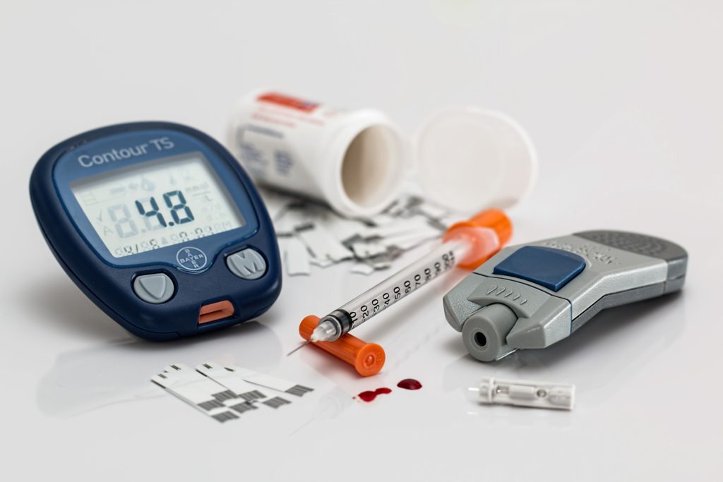
Under normal healthy conditions, the peaks and valleys of insulin release are moderate. Under poor dietary circumstances including ingested sugary drinks, processed carbohydrates or carb only meals, those insulin peaks can be much more extreme.
Here is where monitoring the hormonal effects of food, in particular the Glycemic load, and the timing of those nutrients becomes important. With either an increase in the amount of insulin needed to get the job done or an abrupt saturation of the muscle and liver tissue, the excess glucose has to go somewhere. If you have had poor dietary habits for an extensive period of time, are synthetic exogenous anabolic hormone free, or are not a complete genetic freak, the answer may be between just above your belt line in the form of unsightly love handles or spare tire.
Besides the storage of excess glucose and stimulating cells to use carbohydrates as preferential fuel source, insulin has also been shown to affect fat utilization and mobilization in other ways. It has been shown to decrease the activity of the catecholamines and their ability to bind to adrenergic receptors, leading to a decreased ability to mobilize fat cells.
When the amount of insulin normally required to “get the job done” is not adequate, more and more insulin is required to signal the insulin sensitive receptors on the muscles/liver/fat cells to absorb glucose. If enough insulin is not released from the pancreas, the blood sugar levels stay elevated.
With consistently elevated blood glucose levels, the doors for negative metabolic effects are wide open. Increasing body fat, hypertension, diabetes mellitus, metabolic syndrome, elevated triglycerides, decreased HDL, cognitive decline, as well as many other diseases can quickly arise with insulin resistance.
Did you know that your Suprailiac (think love handles) and Subscapular skinfold sites may reflect insulin sensitivity and blood sugar regulation?
The 1989 Bogalusa Heart Study from The American Journal of Clinical Nutrition found that the suprailliac skinfold was an effective tool in tracking changes in glucose (9). Building on this evidence, a study from the Zaragoza Medical School in Spain looked at the relationship between metabolic complications, in particular insulin dysregulation, and body fat distribution patterning in obese boys. The researchers found significant correlations between the subscapular and suprailliac sites and insulin/blood glucose regulation (Legido 1989).
The researchers also found a correlation between the ratio of subscapular and triceps skinfold measurements and insulin sensitivity. The researchers concluded “an upper central body fat distribution seems to be first associated with an abnormal glucose-insulin homeostasis (13).”
A third study on body fat patterning and insulin, this one from 2006, found a significant correlation between fasting insulin levels and the thickness of the triceps and subscapular skinfolds in 324 healthy young adult north Indian males. The researchers pointed out that the thickness of the subscapular skinfold, in particular, was shown to be a potential predictor of hyperinsulinemia (25).
The skinfold thickness of the sub scapula has also been shown to be better than BMI and WC in identifying hyperinsulinemia in males and females (14).
In a study from Diabetes, a team of researchers measured ten skinfold sites on 7,717 subjects. 360 diabetic and 934 non-diabetic controls were selected. The researchers found that, of the diabetic subjects there was a greater incidence of centripetal fat distribution patterning, which was similar to that of male fat patterning distribution, in both the male and female subjects (7).
The relationship between insulin and other hormones can also have a vast effect on one’s physiology. The balance between insulin, cortisol, and testosterone is an oft-overlooked factor. Especially with regards to human health and fat loss. A disruption in the delicate seesaw between these hormones can lead to a cascade of negative effects, ranging from increased inflammation to metabolic syndrome, as the body is no longer able to adequately fight infection and disease.
In simple terms, when one has increased resistance to insulin, there is typically a disruption in the cortisol/testosterone balance. The increased insulin resistance (a stress on the body) elevates the secretion of the stress hormone, cortisol. Or in some cases vice versa, the increased stress levels signal an increase in cortisol, which then signals the body to go into “storage mode” in order to survive the stress. This storage mode leads to an increase in insulin release.
Estrogen and Stubborn Thigh Fat
Estrogen levels are rising in both humans and animals at alarming rates. Did you know that gynecomastia surgeries are rising nearly every year? Did you ever think we would see the day when manziers, mirdles, and mantyhose would actually exist? How about the fact that beer bellies are beginning to outnumber flat stomachs? Or that hermaphroditic mutated frogs outnumber single sex frogs in certain lakes?
One of the major culprits is the hormone estrogen and its family of estrogen mimicking environmental toxins and chemicals. Estrogen is known as the female sex hormone. Formed from the synthesis of androstenedione from cholesterol, estrogen is formed primarily in the ovaries of women by the stimulating actions of luteinizing hormone (LH) and follicle stimulating hormone (FSH). Estrogen is produced in two forms. First the form known as estradiol, which is formed from testosterone by the enzyme aromatase. The second form known as estrone, is formed from androstenedione.
Androstenedione is one of the precursors to actual testosterone. A male whom does not convert androgens properly to testosterone may have excessive aromatase activity, leading to excessive conversion to estrogen either via the precursor androstenedione or actual testosterone.
Is Estrogen really that bad? Phytoestrogens and Xenoestrogens (environmental estrogen mimickers) have been linked to several cancers as well as growth of tumors. Some of the major problems with estrogen include how and where it is stored, it’s accessibility to the cell membranes, and how easily it can actually be produced from its androgen precursors.
Research on estrogen and fat distribution patterns has been quite revealing. A 1999 study out of Japan looked at the effects menopause had on body fat distribution in 764 subjects. The subjects were broken down into 545 premenopausal and 219 postmenopausal women. Upper and lower body bodyfat levels were measured via DEXA scan. The researchers found that fat accumulated in the lower body is significantly correlated to estrogen hormone concentrations regardless of any increase in overall obesity or aging (12).
A study out of the University of Alabama’s Department of Nutrition Sciences looked at body fat partitioning in fifty-two postmenopausal women. The women had their hormones analyzed and body fat levels measured through DEXA scan. The researchers found a decrease in abdominal fat accumulation in the women undergoing hormone replacement therapy. The downside was, with the increased estrogen, they then began to accumulate more fat around about the thighs (11).
A 2012 study from the Annals of the New York Academy of Sciences found that women who had gone through menopause had 49% more intra-abdominal fat, 36% more fat about their trunk, and 22% more subcutaneous fat about their abdomen (24).
Finally, 1999 research from Italy looked at the fat distribution patterns of 1075 female subjects. These women were divided into one of 4 groups: premenopausal, peri menopausal, postmenopausal, and control. The researchers found significantly less leg fat accumulation in the postmenopausal group, compared to the peri menopausal group. This difference was even more significant when comparing the premenopausal and postmenopausal groups. The major hormonal difference between the two groups: Estrogen Levels (10)!
Testosterone
How important is testosterone to your health? Well, a drop in testosterone has been linked to everything from bone health and low back pain to chronic inflammation and cancer. Testosterone plays a role in keeping energy levels up and inflammation levels down. Beginning in the hypothalamus of the brain, testosterone production is stimulated through a series of hormonal actions. Gonadotropin releasing hormone is secreted from the hypothalamus, which then signals the pituitary gland to secrete Luteinizing hormone. The luteinizing hormone is a signal for the synthesis and release of testosterone.
The trouble with testosterone is that its production is not an easy, one or two step process. Testosterone can be converted into DHT (dihydrotestosterone) or aromatize to estrogen. Two other important factors about testosterone are concentration and sensitivity of the testosterone receptors on tissue and how much testosterone is in the form of free testosterone.
So, how does testosterone and lack thereof affect body fat distribution patterns? A 2001 study from Life Sciences followed male subjects over a six-month period of controlled anabolic steroid administration. The researchers saw a 53% decrease in testosterone, a 77% drop in Luteinizing hormone and an 87% drop in follicle stimulating hormone. To make matters worse, there was a 45% increase in estradiol (2).
The majority of the tissue that became gynecomastia had significant concentration of either androgen or estrogen receptors. The researchers theorized the development of gynecomastia was potentially dependent on three variables:
- Anabolic steroid administration for prolonged periods may cause an excess of circulating estrogens, perhaps through the conversion of testosterone to estrogen;
- A predominant estrogen effect on the breast may produce breast enlargement;
- The presence of estradiol and androgen receptors in gynecomastia tissue suggests that gynecomastia is a hormone dependent event
A separate study found that men with low testosterone stored more fat in their lower body during a six-hour period post-meal. Men with healthy testosterone were burn not only burned more fat during that period of time, but they also had a tendency to store less in their lower body fat depots (20).
Growth Hormone
Known as a peptide hormone, growth hormone is capable of increasing muscle mass, decreasing body fat, healing structural tissue, decreasing inflammation, improving cardiovascular function, basically, fending off the physical symptoms of aging. In excess, though, it can lead to cancer, cardiovascular risks, and diabetes.
Secreted from the anterior pituitary gland, growth hormone production is stimulated by Growth Hormone Releasing Hormone (GHRH) produced in the hypothalamus (somatostatin is the hormone released from the hypothalamus that inhibits growth hormone production). Once growth hormone has been released, it travels to the liver. There it stimulates the liver to secrete a hormone called Insulin like Growth Factor-1 (IGF-1 or somatomedin), which has a similar chemical structure to insulin.
Growth hormone can affect the mobilization of fat cells as GH and IGf-1 receptors are found on the fat cells in your body. When these receptors are activated, they in turn send a signal to begin breakdown of fat so it can be used a fuel.
The body has alpha and beta adrenergic receptors, which are the receptors on the cells responsible for regulation of lipolysis, or fat cell mobilization. They are triggered into action by the catecholamines, epinephrine and norepinephrine. The alpha receptors inhibit fat cell mobilization while the beta receptors stimulate it. Growth hormone stimulates the beta receptors, increasing their responsiveness to the catecholamines.
When it comes to fat accumulation, studies have shown that a decrease in growth hormone can lead to an increase in body fat and obesity. In a 1999 study, researchers looked at the effects discontinuing growth hormone therapy had on body composition in 10 growth hormone deficient subjects. During the initial 3 months of stoppage, the subjects had the greatest increases in subcutaneous fat accumulation. Over the 12-month period, the researchers saw a 48% increase in intra-abdominal fat mass (23).
Another study, this one from 1981, in the American Journal of Clinical Nutrition, found that the knee and calf skinfold measurements were the most effective skinfold sites for determining growth hormone deficiencies in children with growth problems. They also found the greatest discrepancy in the abdomen skinfold measurement site, with the measurement more than double the thickness in growth hormone deficient subjects (21).
Peaking in levels around 3 or 4 am, growth hormone is also released in large amounts after a large meal. Growth hormone production is also stimulated by exercise. Studies have shown that higher intensities and lactic acid production are associated with higher growth hormone output. Studies have shown that when workouts become stale, growth hormone production levels off.
Cortisol
Cortisol is the stress hormone that stimulates our body to go into protection mode during times of crisis. Picture a caveman whom realizes there will be minimal food in the upcoming winter. His body somehow knows he is going to need to store more and more calories to prevent starvation during the cold months ahead. With this perceived stress, his body goes into protection mode, increasing storage of macronutrients in the cells and body tissue. With this increase of macronutrients, more energy is stored and thus weight gain occurs.
An extremely lean caveman would have had a difficult time surviving the winter months with little fat/energy storage to draw upon when the opportunity to hunt/gather presented itself. So in essence, they became fatter due to the effects of cortisol, in conjunction with other hormones such as insulin. Turn the clock forward thousands of years. The body still works the same, but the availability of food is much different. The problem is that stressors are ever-present, whereas back then it may have been intermittent.
Excessive levels of cortisol can then create a viscous cycle, in which people can become more stressed as they begin to gain weight, so they eat more crappy food to spike serotonin to make themselves feel better. With more sugar laden foods, comes more insulin resistance, and of course….more weight gain. Particularly around the stomach area, as numerous studies have found excessive “belly” fat storage is strongly correlated to elevated cortisol levels.
A 1994 study from Obesity Research entitled Stress induced cortisol response and fat distribution in women found a positive correlation between cortisol secretion and abdominal fat distribution (15). A similar study from 1999, this one on men, had similar findings, expressing that cortisol contributed to the subject’s greater abdominal fat depots (5).
A research paper from 2003, associated the stresses of a modern society as contributing factors to the epidemic of abdominal obesity (3). One of the reasons for this cortisol/abdominal obesity link is that visceral fat can have a greater concentration of cortisol receptors than does subcutaneous or brown fat.
Excessive cortisol can lead to the breakdown of muscle proteins into their amino acid constituents, to be converted into usable fuel by the liver. With less muscle comes a metabolic slowdown, and with this metabolic slowdown comes stress, and with greater levels of chronic stress comes elevated cortisol levels, and the cycle begins again.
Too much cortisol can be responsible for many physiological and psychological problems including anxiety, insomnia, depression, cancer, cardiovascular risk factors, obesity, Cushing’s Syndrome, and many other adverse health effects.
Cortisol is not all bad. As a matter of fact, cortisol is the hormone that helps the immune system to fight off inflammation. Kind of ironic. A hormone, when in excess, can cause inflammation, is actually one of the body’s primary defenses against inflammation. Cortisol levels typically peak early in the morning around 7 or 8 am, and drop to a daily low sometime mid-afternoon, roughly around 4pm. It is chronically high cortisol levels we need to worry about.
Thyroid Hormone
According to the textbook, Endocrinology, the authors state “thyroid hormones are extremely important and have diverse actions. They act on virtually every cell in the body to alter gene transcription: under- or over-production of these hormones has potent effects (16)”.
Thyroid hormone, actually there are two, thyroxine (T4) and triiodothyronine (T3), are produced in a small gland at the base of the throat, called the thyroid gland. These hormones are synthesized from the amino acid tyrosine, also responsible for synthesizing the catecholamines, epinephrine and norepinephrine. Thyroxine (T4) is known as the inactive form of thyroid hormone, while triiodothyronine (T3) is the active form. In large concentrations, these hormones can increase the fat burning effects of the catecholamines, as they can increase the number/sensitivity of the Beta-Adrenergic receptors (26).
From controlling your appetite, to maintenance of body temperature and energy levels, the thyroid gland is a major player in your overall well-being. It can affect your sleep patterns, metabolism and sex drive. Thyroid hormone has such an effect on metabolism that bodybuilders and figure athletes have found their way to injectable T3 to increase their metabolism, and thus, burn more body fat.
In healthy individuals, roughly 80% of the thyroid hormone produced is in the “inactive” form of T4, while the other 20% is T3. To become “active”, T4 is stripped of one of its outer ring iodine molecules through a process known as deiodination. Incidentally, the antioxidant/mineral Selenium plays a critical role in this conversion, as the enzyme selenodeiodinase is responsible for stripping the iodine molecule from the outer ring of T4.
Without proper Selenium levels, this process may become irregular, potentially leading to a cascade of negative events, the least of which is a drop in your metabolic rate. A deficiency in iodine may have similar effects as this is also an important component in the synthesis of thyroid hormones.
Speaking of slowing down metabolic rate, the stress hormone cortisol can affect thyroid production in two ways,
- Cortisol has receptors on the pituitary gland that can shut down the production of TSH, thyroid stimulating hormone, which basically relays the message from the hypothalamus to the thyroid gland, to produce thyroid hormones.
- Cortisol can affect the conversion of “inactive” T4 to “active” T3. Corticosteroids and dietary induced cortisol elevations can affect the 5-deiodinase enzyme, which is responsible for converting up to one third of the T4 to T3. With the impairment of this enzyme, an iodine molecule from the inner ring may be taken instead. This leads to an inactive form of T3, known as reverseT3.
Estrogen can affect thyroid hormone as it can increase the amount of TBG, Thyroxine Binding Globulin. When a hormone is “bound” to one of these binding globulins, it may become unavailable for uptake into its receptor cells. Certain foods, especially certain vegetables when eaten uncooked, can have negative effects on one’s thyroid functioning. These are known as goitrogens.
Soy, stress, and lack of certain minerals such as zinc and magnesium have also been shown to dramatically effect thyroid functioning. It is theorized that stress can signal the brain to turn down production of TRH and TSH, potentially leading to hypothyroidism.
The Catecholamines
The hormones known as the Catecholamines (epinephrine/adrenaline, norepinephrine/noradrenaline, and dopamine) are similar to growth hormone in that these are also formed from amino acids, specifically Tyrosine and its derivatives. Unlike growth hormone, the catecholamines are catabolic in nature. They trigger the breakdown of tissue/cells to be used as fuel.
Synthesis of catecholamines begins with the amino acid tyrosine. Derived from the amino acid phenylalanine, Tyrosine (from the Greek word “tyros” for cheese as it is found in casein protein) is responsible for the synthesis of the catecholamines, as well as other hormones including thyroid and adrenal hormones.
Dopamine is the precursor to norepinephrine, which is the precursor to epinephrine. Tyrosine is responsible for the synthesis of dopamine, so without the amino acid tyrosine, catecholamine production may be interrupted.
Dopamine the plays a key role in several functions of human health including movement, mood, nervous system activation, pain perception, and especially brain function with regards to reward/motivation systems. Parkinson’s Disease and the neurological symptoms associated with it are linked to depressed dopamine levels. Addiction is another dopamine related health consequence, as the ingested drugs can increase dopamine levels, putting the user in a euphoric state, but then comes the crash, and the only way to fend off the rebound effects is to raise dopamine levels again by, you guessed it, taking more drugs.
Secreted from the adrenal medulla, epinephrine is part of the endocrine system. The sympathetic nervous system, which is responsible for the fight or flight response, is where the catecholamine norepinephrine exerts its effects, while the endocrine system is the major stomping ground epinephrine.
From increased blood flow to the working muscles, to decreased blood flow to the digestive system, we have all felt the effects of the catecholamines. Increases in heart rate, body temperature, and even that “adrenaline” rush is a response to the effects of the catecholamines.
Catecholamines also play a role in thermogenesis. They have a receptor on BAT cells, which when activated, turns on the body’s ability to produce heat and stimulate metabolism.
Leptin
Produced in the White Adipose Tissue (WAT), along with another hormone called resistin, leptin plays a critical role in appetite, hunger and satiety. It lets you know when you are full. Leptin is similar to other hormones in that it circulates through the bloodstream and binds to receptor sites, yet it is different in that it is synthesized in the fat cells themselves.
In the brain, leptin influences the actions of Neuropeptide Y (NPY), an appetite regulating neurotransmitter that signals the brain to decrease metabolic rate or increase body fat storage. If levels of this NPY are elevated, overeating and body fat accumulation may result. With low levels of Leptin, NPY levels are allowed to elevate and vice versa.
In obesity, the sensitivity of the leptin receptors can decrease, requiring more leptin to do the job. Eventually the effectiveness of the high concentrations of leptin is not enough and the system begins to break down.
It has been shown that the obesity can lead to leptin resistance, thus creating a viscous cycle for weigh gain and fat accumulation. The more obese you become, the more fat cells you have. The more fat cells you have, the more leptin you are capable of producing. The more leptin you can produce; the more leptin resistant your leptin receptors can become. NPY levels are then elevated, and you eat more.
Leptin also plays a role in the regulation of other hormones. The ripple effect of compromised leptin levels can be seen throughout the body. For instance, leptin regulates NPY, which when left unchecked can increase the production of cortisol in the adrenal glands, through its actions on Corticotrophin-releasing hormone (CRH) in the hypothalamus.
Neuropeptide Y
This neurotransmitter is linked to many functions in the brain, including cognitive functioning, energy, and appetite regulation and satiety. With regards to fat loss/accumulation, its actions include signaling the body to store more fat and increase ingestion of food/calories. High stress and ingestion of highly processed food can stimulate the release of NPY.
Ghrelin
Ghrelin is a hormone that signals the body when it is hungry, increasing prior to a meal and dropping after.
Glucagon
Glucagon is a digestive hormone, released when food, particularly protein, leaves the stomach and enters the small intestine. It slows the exit of food from the stomach, making you feel full; acts to raise blood sugar, and tells the liver to pump out glucose. It is one of a number of counter-insulin hormones, including cortisol, GH, and epinephrine.
Question
How do we lose this fat? Is spot reduction possible? What are some training, nutrition, detox, supplementation, and health hacks/strategies to fight back against stubborn fat and finally win the battle of the bulge?
Stay tuned for Part 2 of Problem Area Fat Loss (2021). If you are interested in much more on this topic, be sure to pick up a copy of our book Butts, Guts, and Other Problem Areas on Amazon.com today!
Thx for reading!
References
- Albrink M, Meigs W. Interrelationship between skinfold thickness, serum lipids and blood sugar in normal men. The American Journal of Clinical Nutrition. 15(5); Pp 255-261. 1964
- Calzada L, Torres-Calleia J, Martinez J, Pedron N. Measurement of androgen and estrogen receptors in breast tissue from subjects with anabolic steroid-dependent gynecomastia. Life Sciences. 69(13); Pp 1465-1469. 2001.
- Drapeau V, Therrien F, Richard D, Tremblay A. Is visceral obesity a physiological adaptation to stress? Panminerva Medica. 45(3); Pp 189-195. 2003.
- Ebbeling C, Leidig M, Feldman H, Lovesky M, Ludwig D. Effects of a low-glycemic load vs low-fat diet in obese young adults: a randomized trial. JAMA. 297(19); Pp 2092-2102. 2007.
- Epel E, Moyer A, Martin C, Macary S, Cummings N, Rodin J, Rebuffe-Scrive M. Stress-induced cortisol, mood, and fat distribution in men. Obesity Research. 7(1); Pp 9-15. 1999.
- Eston L. Changes in performance, skinfold thickness, and fat patterning after three years of intense athletic conditioning in high level runners. British Journal of Sports Medicine. 39; Pp 851-856. 2005.
- Feldman R, Sender J, Siegelaub A. Difference in diabetic and nondiabetic fat distribution patterns by skinfold measurement. Diabetes. 18(7); Pp 487-486. 1969.
- Foster M, Pagliassotti M. Metabolic alterations following visceral fat removal and expansion: Beyond anatomic location. Adipocyte. 1(4); Pp 192-199. 2012.
- Freedman D, Srinivasan S, Harsha D, Webber L, Berenson G. Relation of body fat patterning to lipid and lipoprotein concentrations in children and adolescents: the Bogalusa Heart Study. American Journal of Clinical Nutrition. 50; Pp 930-939. 1989.
- Gambacciani M, Ciaponi M, Cappagli B, Benussi C, De Simone L, Genazzani A. Climacteric modifications in body weight and fat tissue distribution. Climacteric. 2(1); Pp 37-44. 1999.
- Goss A, Darnell BE, Brown M, Oster R, Gower B. Longitudinal associations of the endocrine environment on fat partitioning in postmenopausal women. Obesity (Silver Spring). 20(5); Pp 939-944. 2012.
- Ijuin H, Douchi T, Oki T, Maruta K, Nagata Y. The contribution of menopause to changes in body-fat distribution. J Obstet Gynaecol Res. 25(5); Pp 367-372. 1999.
- Legido A, Sarria A, Bueno M, Garagorri J, Fleta J, Ramos F, Abos M, Perez-Gonzalez J. Relationship of body fat distribution to metabolic complications in obese prepubertal boys: gender related differences. Acta-Pediatrica Scandanavia. 78(3); Pp 440-446. 1989.
- Misra A, Madhavan M, Vikram N, Pandey R, Dhingra V, Luthra K. Simple anthropometric measures identify fasting hyperinsulinemia and clustering of cardiovascular risk factors in Asian Indian adolescents. Metabolism. 55(12); Pp 1569-1573. 2006.
- Moyer A, Rodin J, Grilo C, Cummings N, Larson L, Rebuffé-Scrive M. Stress-induced cortisol response and fat distribution in women. Obesity Research. 2(3); Pp 255-262. 1994
- Nussey S, Whitehead S. Endocrinology: an integrated approach. Informa Healthcare. 2001.
- Rosenbaum M, Prieto V, Hellmer J, Boschmann M, Krueger J, Leibel RL, Ship A. An exploratory investigation of the morphology and biochemistry of cellulite. Plastic and Reconstructive Surgery. 101; Pp 1934-1939. 1998.
- Rosenow A et all. Identification of Novel Human Adipocyte Secreted Proteins by Using SGBS Cells. Journal of Proteome Research. 9(10); Pp 5389-5401. 2010.
- Rossi A, Vergnanini A. Cellulite: a review. Journal of the European Academy of Dermatology & Venereology. 14; Pp 251-262. 2000.
- Santosa S, Jensen M. Effects of male hypogonadism on regional adipose tissue fatty acid storage and lipogenic proteins. PLoS One. 7(2); e31473. 2012.
- Satinder B, Moffitt S, Goldsmith M, Bain R, Kutner M, Rudman D. A method for screening growth hormone deficiency using anthropometrics. American Journal of Clinical Nutrition. 34(2); Pp 281-288. 1981.
- Seltzer C, Mayer J. Greater reliability of the triceps skin fold over the subscapular skin fold as an index of obesity. The American Journal of Clinical Nutrition. 20(9); Pp 950-953.1967.
- Stouthart P, de Ridder C, Rekers-Mobarg L, van der Waal H. Changes in body composition during 12 months after discontinuation of growth hormone therapy in young adults with growth hormone deficiency from childhood. Journal of Pediatric Endocrinology and Metabolism. 12(1); Pp 335-338. 1999.
- Toth M, Tchernof A, Sites C, Poehlman E. Menopause-related changes in body fat distribution. Ann N Y Acad Sci. 904; Pp 502-506. 2000.
- Vikram N, Misra A, Pandey R, Dwiyedi M, Luthra K, Dhingra V, Talwar K. Association between subclinical inflammation and fasting insulin in urban young adult north Indian males. The Indian Journal of Medical Research. 124(6); Pp 677-682. 2006.
- Wahrenberg H, Engfeldt P, Arner P, Wennlund A, Ostman J. Adrenergic regulation of lipolysis in human adipocytes: findings in hyper- and hypothyroidism. Journal of Clinical Endocrinology and Metabolism. 63(3); Pp 631-638. 1986.
